
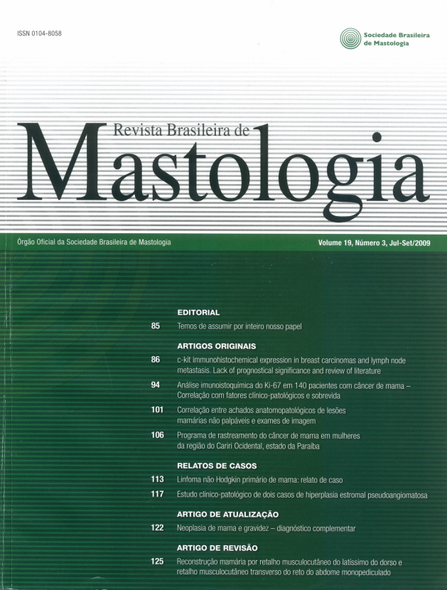
Correlation between anatomopathological real find of non-palpable breast lesions and test of image
Keywords:
Stereotactic localization, Benign lesions, Breast cancerAbstract
Objectives: To describe surgical biopsies after wire localization of non palpable mammary lesions and
to consider their anatomopathological correlation. Methods: A descriptive and retrospective study was
realized through analysis of handbooks of patients at Mastology Clinics of the Instituto de Previdência
dos Servidores do Estado de Minas Gerais (IPSEMG). All cases ofpatients submitted to surgical biopsy of
non-palpable mammary lesion after wire localization for mammography or ultrasound, during the period
of 8/3/2004 to 5/25/2006, were selected. The variables analyzed were: age ofthe patient at the time of the
surgery, type of breast lesion presented during the tests of image and anatomopathological result. Results:
There were a total of 261 surgeries, which 236 cases were elected. The average age of the patients was 50,9
years (19-91 years). In 120 cases (50.85%) the biopsy was made due to the mammary microcalcifications;
in 95 cases (40.25%) for nodule; in 15 cases (6.36%) forfocal asymmetry and in 6 cases (2.54%) for architectural distortion. The anatomopathological result was benign in 170 cases (72.03%) and malignantin 66 (27.97%). Conclusions: The casuistic of the Mastology Service of IPSEMG is compatible with the
data founded in literature. The surgical management of non-palpable breast lesions after wire localization
is a procedure which is able to establish the differential diagnostic of these lesions, breast cancer diagnosis in
early stage and to promote the complete removal ofthe injuries in frequently single surgical time.
Downloads
References
1.
2.
3.
4.
5.
6.
Instituto Nacional do Câncer (Inca). Estimativa da incidência e mortalidade por câncer no Brasil. Brasília (DF): Ministério da Saúde; 2008. Disponível em: http:// www.inca.gov.br/
estimativa/2008.
Silverstein MJ, Gamagami P, Rosser RJ, Gierson ED, Colburn
WL, Handel N, et al. Hooked-wire directed breast cancer biopsy
and overpenetrated mammography. Cancer. 1987;59(4):715-22.
Pinotti JA. Abordagem do carcinoma não palpável em tempo cirúrgico único. In: Câncer de mama inicial: aspectos atatuais. São
Paulo: Fundo Editorial BYK; 2003. p. 98-105.
Valejo FAM, Tiezzi DG, Campos DA, Felix FL. Avaliação da eficiência da biópsia excisional após agulhamento de lesões não palpáveis
da mama. Rev Bras Ginecol Obstet. 2007;29(4):200-04.
Buijs-Van Der Woude T, Verkooijen HM, Pijnappel RM, Klinkenbijl JH, Rinkes IH, Peeters PH, et al. Cost comparison between
stereotactic large-core-needle biopsy versus surgical excision biopsy in the Netherlands. Eur J Cancer. 2001;37(14):1736-45.
Boener S, Fornage BD, Singletary E, Sneige N. Ultrasound-guided
fine-needle aspiration (FNA) of nonpalpable breast lesion: a review
of 1885 FNA cases using the National Cancer Institute - supported recommendations on the uniform approach to breast FNA.
Cancer. 1999;87(1):19-24.7. Menke CH, Biazus JV, Xavier NL, Cavalheiro JA, Rabin EG, Cericatto R, et al. Conduta nas lesões mamárias não palpáveis. In: Rotinas em Mastologia. 2ª ed. Porto Alegre: Artmed; 2007. p. 75-87.
8. Papatestas AE, Hermann D, Hermann G, Tsevdos C, Lesnick G. Surgery for nonpalpable breast lesions. Arch Surg. 1990;125(3):399-402.
9. Markopoulos C, Kakisis J, Kouskus S, Kontzoglou K, Koufopoulos K, Gajas G. Management of nonpalpable, mammografically
detectable breast lesions. World J Surg. 1999;23(5):434-8.
10. Ernst MF, Avenarius JKA, Schuur KH, Roukema JA. Wire localization of non-palpable breast lesions: out of date? Breast.
2002;11(5):408-13.
11. Melhado VC, Alvares BR, Almeida OJ. Correlação radiológica е
histológica de lesóes mamárias não palpáveis em pacientes submetidas a marcação pré-cirúrgica, utilizando-se o sistema BI-RADS.
Radiol Bras. 2007;40(1):9-11.12. Markopoulos C, Kouskus E, Gogas H, Mandas D, Kakisis J, Gogas J. Factors affecting axillary lymph node metastases in patients
T1 breast carcinoma. Am Surg. 2000;66(11):1011-3.
13. Pijnappel RM, Peeters PHM, Van Den Donk M, Holland R, Hendricks JHCL, Deurloo EE, et al. Diagnostic strategies in non-palpable breast lesions. Eur J Cancer. 2002;38(4):550-5.
14. Salles MA, Matias MARF, Perez AA, Gobbi H. Carcinoma ductal in situ da mama: critérios para diagnóstico e abordagem em
hospitais públicos de Belo Horizonte. Rev Bras Ginecol Obstet.
2006;28(12):721-7.
15. Abrahamson PE, Dunlap LA, Amamoo MA, Schell MJ, Braeuning
MP, Pisano ED. Factors predicting succesfull needle-localizated
breast biopsy. Acad Radiol. 2003;10(6):601-6.
16. Ernst MF, Roukema JA. Diagnosis of non-palpable breast cancer: a
review. Breast. 2002;11(1):13-22.
Downloads
Published
How to Cite
Issue
Section
License
Copyright (c) 2026 Cássio Furtini Haddad, Fernanda Miranda dos Reis, Valéria Cristina Pereira Almeida, Frederico Viana Negrão de Lima, Edite de Fátima Pinto, Cristóvão Pinheiro Barros, Annamaria Massahud Rodrigues dos Santos

This work is licensed under a Creative Commons Attribution 4.0 International License.