
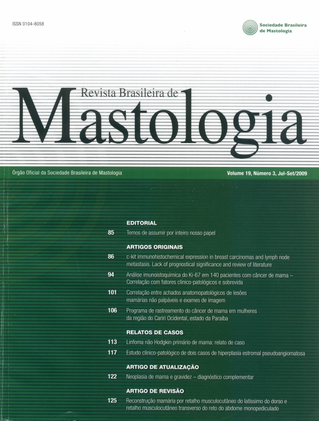
c-kit immunohistochemical expression in breast carcinomas and lymph node metastasis. Lack of prognostical significance and review of literature
Keywords:
c-kit, Breast carcinomas, ImmunohistochemistryAbstract
Objective: To evaluate the expression of c-kit in breast invasive ductal carcinomas (IDC) and metastasis to lymph nodes considering epithelial and stromal components separately and correlate
this variable to others clinical, pathological and biological markers (EGFR, Her-2, ER, PR, Ki-67
and p53 expression). Methods: We analysed 80 IDC, stage T2-T4 Nx Mx, in TMA of epithelial, stromal component and lymph nodes. Statistical analysis considered significant a p value
< 5%. Results: c-kit expression was founded in 9 cases in epithelial component (11.3%) and in 10 cases (12.5%) of stromal component. The 43 samples of lymph nodes metastasis were negative. EGFR
and Her-2 were predominantly negative, both in epithelial (77.5% and 73.75%, respectively), as stromal (97.5% and 95.0%) components and metastasis to lymph nodes (83.7% and 62.8%). While
ER, PR, Ki-67 and p53 were positive in 49 (61.0%), 40 (53.0%), 67 (83.75%) and 59 cases (73.75%)
in the epithelial component. Stromal cells have proved negatives. c-kit epithelial expression correlated
to presence of in situ component (p = 0.044) and stromal c-kit expression correlated to presence of
necrosis (p = 0.002). There was no association between c-kit expression and staging and biological
markers. Transformed epithelial cells at the lymph nodes metastasis stained for ER, PR, Ki-67 and
p53 in 27 (62.8%), 16 (37.5%), 41 (95.0%) and 28 cases (65.1%), respectively. Conclusions: The expression of c-kit is mostly negative in primary IDC both in the epithelial and stromal component,
as well as in lymph node metastasis. The lack of correlation between c-kit and others tyrosine kinase
proteins suggest that they are independently regulated. Metastasis for lymph nodes were not c-kit positive and further studies of mutations of c-kit and his family, correlate with other prognostic factors
and survival required to reveal the exact mechanism of action of this molecule in breast cancer.
Downloads
References
1. Ross JS,Fletcher JA. Targeted therapy in breast cancer. The HER-2/
neu gene and protein. Mol Cell Proteomics. 2004;3(4):379-98.
2. Perou CM, Sorlie T, Eisen MB, Van de Rijn M, Jeffrey SS, Rees
CA, et al. Molecular portraits of human breast tumours. Nature.
2000;406:747-52.
3. DeMatteo RP, Heinrich MC, El-Rifai WM, Demetri G. Clinical
management of gastrointestinal stromal tumors: before and after
STI571. Hum Pathol. 2002;33(5):466-77.
4. Akin C, Metcalfe DD. The biology of Kit in disease and the
application of pharmacogenetics. J Allergy Clin Immunol.
2004;114(1):13-9; quiz 20.
5.
6.
7.
Diallo R, Rody A, Jackisch C, Tinge E, et al. C-KIT expression in
ductal carcinoma in situ of the breast: co-expression with HER-2/
neu. Hum Pathol. 2006;37:205-11.
Cohen PS, Chan JP, Lipkunskaya M, Biedler JL, Seeger RC. Expression of stem cell factor and c-kit in human neuroblastoma. The
Children's Cancer Group. Blood. 1994;84(10):3465-72.
Hines SJ, Organ C, Kornstein MJ, Krystal GW. Coexpression of
the c-kit and stem cell factor genes in breast carcinomas. Cell Growth Differ. 1995;(6):769-79.
8. Joensuu H, Puputti M, Tynninen O, Sihto H, Blom T, Mäenpää
H, et al. Amplification of KIT, PDGFRA, VEGFR2, and EGFR
in gliomas. Mol Cancer Res. 2006;4(12):927-34.
9. Nalwoga H, Arnes JB, Wabinga H, Akslen LA. Expression of
EGFR and c-kit is associated with the basal-like phenotype in breast carcinomas of African women. APMIS. 2008;116(6):515-25.
10. Simon R, Panussis S, Maurer R, et al. KIT (CD117)-positive breast
cancers are infrequent and lack KIT gene mutations. Clin Cancer
Res. 2004;10:178-83.
11. Azoulay S, Laé M, Fréneaux P, Merle S, Al Ghuzlan A, Chnecker
C, et al. KIT is highly expressed in adenoid cystic carcinoma of the
breast, a basal-like carcinoma associated with a favorable outcome.
Mod Pathol. 2005;18(12):1623-31.
12. Roussidis AE, Theocharis AD, Tzanakakis GN, Karamanos
NK. The importance of c-Kit and PDGF receptors as potential
targets for molecular therapy in breast cancer. Curr Med Chem.
2007;14(7):735-43.
13. Cristofanilli M, Morandi P, Krishnamurthy S, Reuben JM, Lee
BN, Francis D, et al. Imatinib mesylate (Gleevec) in advanced breast cancer-expressing C-Kit or PDGFR-beta: clinical activity and
biological correlations. Ann Oncol. 2008;19(10):1713-9.
14. Hornick JL, Fletcher CD. Immunohistochemical staining for KIT
(CD117) in soft tissue sarcomas is very limited in distribution. Am
J Clin Pathol. 2002;117:188-93.
15. Ko CD, Son BH, Ahn SH, Ka IW, Gong GY, Kim JC. Significance of mismatch repair protein expression in the chemotherapeutic
response of sporadic invasive ductal carcinoma of the breast. Breast
Journal. 2004;10(1):20-6.
16. Tsuda H, Morita D, Kimura M, Shinto E, Ohtsuka Y, Matsubara
O, et al. Correlation of KIT and EGFR overexpression with invasive ductal breast carcinoma of the solid-tubular subtype, nuclear
grade 3, and mesenchymal ormyoepithelial differentiation. Cancer
Sci. 2005;96:48-53.
17. Allred DG, Harvey JM, Berardo M, Clark GM. Estrogen receptor
status by immunohistochemistry is superior to the ligand-binding
assay for predicting response do adjuvant endocrine therapy in breast cancer. J Clin Oncol. 1999;17:1474-81.18. Bankfalvi A, Boecker W, Reiner A. Comparison of automated and
manual determination of HER2 status in breast cancer for diagnostic use: a comparative methodological study using the Ventana BenchMark automated staining system and manual tests. Int J
Oncol. 2004;25(4):929-35.
19. Tsutsui S, Yasuda K, Suzuki K, Takeuchi H, Nishizaki T, Higashi H, et al. A loss of c-kit expression is associated with an advanced stage and poor prognosis in breast cancer. Br J Cancer. 2006;94:1874-8.
20. Natali PG, Nicotra MR, Sures I, Mottolese M, Botti, C, Ullrich A.
Breast cancer is associated with loss of the c-kit oncogene product.
Int J Cancer. 1992;52:713-7.
21. Matsuda R, Takahashi T, Nakamura S, Sekido Y, et al. Expression of the c-kit protein in human solid tumors and in corresponding fetal and adult normal tissues. Am J Pathol. 1993;
142:339-46.
22. Chui X, Egami H, Yamashita J, et al. Immunohistochemical expression of the c-KIT proto-oncogene product in human malignant
and non-malignant breast tissues. Br J Cancer. 1996;73:1233-6.
23. Tsuura Y, Suzuki T, Honma K, Sano M. Expression of c-KIT protein in proliferative lesions of human breast: sexual difference and
close association with phosphotyrosine status. J Cancer Res Clin
Oncol. 2002;128:239-46.
24. Palmu S, Söderström KO, Quazi K, Isola J, Salminen E. Expression of c-KIT and HER-2 tyrosine kinase receptors in poor-prognosis breast cancer. Anticancer Res. 2002;22(1A):411-4.
25. Ko CD, Kim JS, Ko BG, et al. The meaning of the c-KIT protooncogene product in malignant transformation in human mammary epithelium. Clin Exp Metastasis. 2003;20:593-7.
26. Yared MA, Middleton LP, Bernstam FM, Cristofanilli M, Sahin
AA. Expression of c-kit proto-oncogene product in breast tissue.
Breast J. 2004;10(4):323-7.
27. Ulivi P, Zoli W, Medri L, et al. C-KIT and SCF expression in normal
and tumor breast tissue. Breast Cancer Res Treat. 2004:83:33-42.
28. Maffini MV, Soto AM, Sonnenschein C, Papadopoulos N, Theoharides TC. Lack of c-kit receptor promotes mammary tumors in Nnitrosomethylurea-treated Ws/Ws rats. Cancer Cell Int. 2008;8:5.
29. Polat А. с-KIT еxpression in columnar cell lesions of the breast
accompanied by benign and malignant breast diseases. Pathol Res
Pract. 2007;203:765-9.
30. Djordjevic B, Hanna WM. Expression of c-kit in fibroepithelial lesions of the breast is a mast cell phenomenon. Mod Pathol.
2008;21(10):1238-45.
31. Tan PH, Jayabaskar T, Yip G, Tan Y, Hilmy M, Selvarajan S, et al.
p53 and c-kit (CD117) protein expression as prognostic indicators
in breast phyllodes tumors: a tissue microarray study. Mod Pathol.
2005;18(12):1527-34.
32. Kononen J, et al. Tissue microarrays for high-throughput molecular profiling of tumor specimens. Nat Med. 1998;4(7):844-7.
33. Camp RL, Charette LA, Rimm DL, et al. Validation of tissue
microarray technology in breast carcinoma. Lab Invest. 2000;
80(12):1943-9.
34. Zhang D, et al. Reliability of tissue microarrays in detecting protein expression and gene amplification in breast cancer. Mod Pathol. 2003;16(1):79-84.
35. Loring P, et al. HER2 positivity in breast carcinoma: a comparison of chromogenic in situ hybridization with fluorescence in situ
hybridization in tissue microarrays with targeted evaluation of intratumoral heterogeneity by in situ hybridization. Appl Immunohistochem Mol Morphol. 2005;13(2):194-200.
36. Henriksen KL, et al. Semi-quantitative scoring of potentially predictive markers for endocrine treatment of breast cancer: A comparison between whole sections and tissue microarrays. J Clin Pathol.
2006;14-8.
37. Logullo AF, Godoy AB, Mourão-Neto M, Simpson AJ, Nishimoto
IN, Brentani MM. Presence of ductal carcinoma in situ confers
an improved prognosis for patients with T1NOM0 invasive breast
carcinoma. Braz J Med Biol Res. 2002;35(8):913-9.
38. Selvarajan S, et al. c-erbB-2 (HER-2/neu) immunohistochemistry
in invasive breast cancer: is there concordance between standard
sections and tissue microarrays? Pathology. 2006;38(4):316-20.
39. Cristofanilli M, Morandi P, Krishnamurthy S, Reuben JM, Lee
BN, Francis D, et al. PDGFR-beta: clinical activity and biological
correlations. Ann Oncol. 2008;19(10):1713-9.
Downloads
Published
How to Cite
Issue
Section
License
Copyright (c) 2026 Fiorita G. L. Mundim, Angela F. Logullo, Miriam F. B. Engelman, Suely Nonogaki, Luciana Logullo, Fernando Augusto Soares

This work is licensed under a Creative Commons Attribution 4.0 International License.