
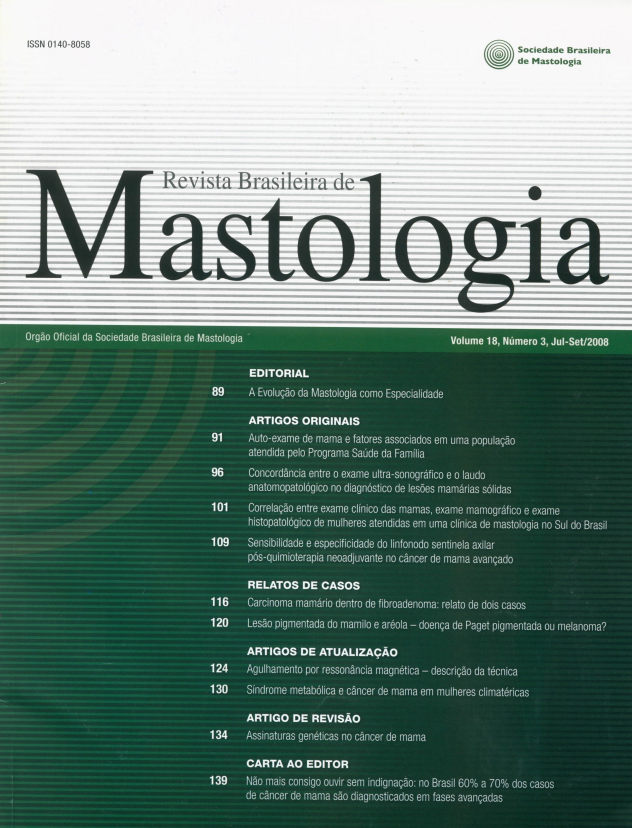
Correlation between clinical breast examination, mammography and histopathology examination of women attending in a mastology clinic at the South of Brazil
Keywords:
Cancer, linical and mammographyAbstract
Objective: The aim of the present study was to correlate the clinical breast examination, mammography and histopathology, to verify the sensitivity, specificity, negative and positive predictive
values. Materials and Methods: Were analized 193 of pacientes seen at a private clinic in the
city of Santo Angelo, RS. The variables identified were the pacients' age, the clinical exam results,mammography and histopathological. Results: The average of the women was 48.5 years, varying
from 18 to 92. 55.4% had negative exam or benignant alteration on the histopathological. When
the histopathological was correlated with the clinical breast examination, a significant association
was seen (p < 0.001), with sensitivity 94.2%, specificity 33.6%, negative and positive values
87.8% and 53.3%. The histopathological shows bigger corralation inside BI-RADS V, with sensitivity 54.7%, specificity 99.1%, negative and positive values 73.1% and 97.9%. Conclusion:
Mammary touching alone can be a simple, fast and efficient screening clinical examination for
breast cancer. The histopathological shows bigger correlation inside BI-RADS * 0, I, II, III. Рacients with BI-RADS * IV, V must be submitted to histopathological exam for the high risk of
associated malignant disease.
Downloads
References
1. Aguillar VLM, Bauab SP. Rastreamento mamográfico para detecção precoce do câncer de mama. RBM. 2003;13:82-9.
2. Fernandes SS, Pereira ASP, Montenegro CAB, Koch HA, Lima
ML. Ultra-sonografia tridimensional da mama: uma expectativa.
RBM. 2004;14:91-4.
4. Brasil. Ministério da Saúde (MS). Instituto Nacional do Câncer
(INCA). Estimativas 2008. Incidência de Câncer no Brasil. [Acessado em 1 jul 2008]. Disponível em <http://www.inca.gov.br>.
5. Harris JR, Lippman ME, Morrow M, Osborne CK. Diseases ofthe
breast. 3. ed. Philadelphia; Lippincott Williams & Wilkins, 2004.
p.133-6.
3. Menke CH, Biazús JV, Xavier NL, et al. Rotinas em Mastologia 2.
ed. Porto Alegre; Artmed. 2007. p.129-31.
6. Parking DM, Malone KE, Porter PL, et al. Global cancer statistics.
Cancer J Clin. 2005;55:74-108.
7.
8.
Godinho ER, Koch HA. Rastreamento do câncer de mama: aspectos relacionados ao médico. Radiol Bras. 2004:37:91-9.
Borghesan DHP, Baraúna M, Pelloso SM. Auto-exame das mamas: conhecimento e prática entre profissionais da área da saúde de
uma instituição pública / Self-examination of breast: knowledge
and practice in government health professionals. Acta Sci Health
Sci. 2003: 25:103-13.
9. Marinho LAB, Costa-Gurgel MS, Cecatti JG, Osis MJD. Conhecimento, atitude e prática do autoexame das mamas em centros de
saúde. Rev Saúde Pública. 2003;37(5):576-82.
10. Molina L, Dalben I, De Luca LA. Análise das oportunidades de
diagnóstico precoce para as neoplasias malignas de mama. Rev Assoc Med. Bras. 2003;49:185-90.
11.
12.
13.
Brasil. Ministério da Saúde. Instituto Nacional do Câncer (INCA).
Controle do câncer de mama: Documento de consenso. 2004.
[Acesso em: 20 ago 07]. Disponível em: .
Pfarl G, Helbich MD TH; BI-RADS®. Breast Imaging Reporting
and Data System. 4. ed. Copyright 1992, 1993, 1995, 1998, 2003
American College of Radiology. [Acesso em: 20 ago 2007]. Disponível em: <http://www.birads.at>.
Wong AY, Salisbury E, Bilous M. Recent developments in stereotactic breast biopsy methodologies: an apdade for the surgical pathologist. Adv Anat Pathol. 2000;7:6-35.15. Kolb TM, Llchy J, Newhouse JH. Comparison of the performance
of screening mammography, physical examination, and breast US
and evaluation of factors that influence them: an analysis of 27,825
patient evaluations. Radiology. 2002;225:165-75.
14. Menke CH, Biazús JV, Xavier NL, et al. Rotinas em Mastologia 1.
ed. Porto Alegre. Artmed. 2005. p.99-102.
16. Moraes AB, Zanini RR, Turchiello MS, Riboldi J, Medeiros LR.
Estudo da sobrevida de pacientes com câncer de mama atendidas
no hospital da Universidade Federal de Santa Maria, RS, Brasil.
Cad Saúde Pública. 2006;22(10):2219-28.
17. Urban LA. Biópsia percutânea a vácuo (mamotonia) guiado por ultra-sonografia: experiência de 404 casos. Dissertação apresentada
como requisito parcial à obtenção do grau de Mestre. Universidade
Federal do Parana, Curitiba, 2005. p.1-125.
18. Mahoney L, Csima A. Efficiency of palpation in clinical detection
of breast cancer. CMA Toronto J. 1982;127(8):729-31.
19. Yang WT, Tse GMK. Sonographic, mommographic, and histopathologic correlation of symptomatic ductal carcinoma in situ. Am
Roentgen Ray Soc. 2004;182:101-10.
20. Vieira AV, Toigo FT. Predição de malignidade em pacientes das
categorias 4 e 5 BI-RADS; Radiol Bras. 2004;37:25-7.
21. Scaranelo AM, Barros N. Normatização no laudo de mamografia
no Brasil: a utilização do modelo americano (BI-RADS) também
na clínica privada. Radio Brás. 2000;33:311-6.
Downloads
Published
How to Cite
Issue
Section
License
Copyright (c) 2026 Rômulo Andrade de Vargas, Edison Barbosa de Vargas, Vera Regina Andrade Vargas, Honório Sampaio Menezes

This work is licensed under a Creative Commons Attribution 4.0 International License.