
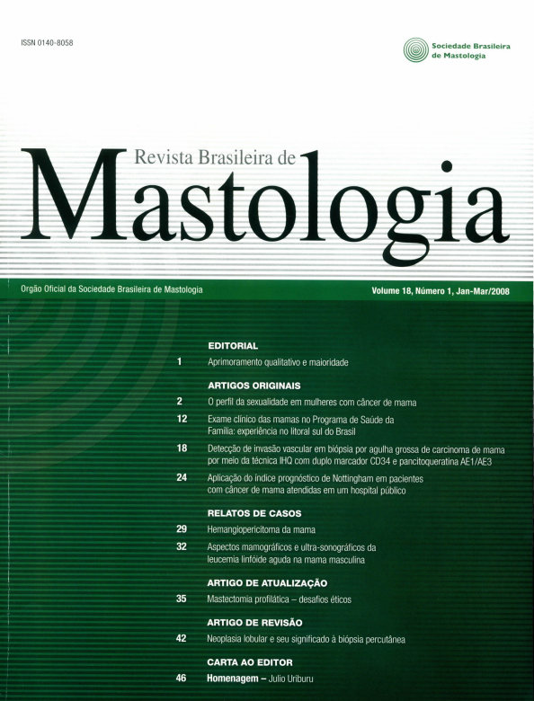
Detecção de invasão vascular em biópsia por agulha grossa de carcinoma de mama por meio da técnica IHQ com duplo marcador CD34 e pancitoqueratina AE1/AE3
Detection of blood vessel invasion in breast cancer core-biopsy through immunohistochemical analysis with double staining CD34 and pan cytokeratin AE1/AE3
Palavras-chave:
Câncer de mama, Invasão vascular, IHQ (imunoistoquímica).Resumo
Objetivos: Detectar a presença de invasão vascular nas biópsias por agulha grossa de carcinoma de
mama por meio da técnica IHQ (IHQ) com duplo marcador (CD34-pancitoqueratina AE1/AE3);
analisar a sensibilidade, a especificidade, o valor preditivo positivo (VPP) e o valor preditivo negativo
(VPN) com essa técnica e comparar a acurácia do duplo marcador com a técnica da hematoxilinaeosina (H&E). Métodos: Cortes histológicos de 74 casos de biópsias por agulha grossa de pacientes
com câncer de mama foram submetidos à coloração por IHQ com duplo marcador e coloração por
H&E. Como grupo-controle, utilizou-se os resultados anatomopatológicos das cirurgias. Resultados:
Invasão vascular ocorreu em 34 casos (45,9%). O teste H&E demonstrou valores superiores à IHQ
quanto a sensibilidade, especificidade, VPP e VPN. Apenas em casos sem metástases axilares, a especificidade foi superior no teste com IHQ. Conclusão: O teste com maior acurácia para a detecção de
invasão vascular nas biópsias foi o da coloração por H&E. Nos casos sem metástases axilares, o duplo
marcador mostrou-se superior somente quanto à especificidade. Por causa da escassez de material das
biópsias, a principal suposição é que, no material proveniente das cirurgias, que é mais representativo,
ou em biópsias com maior número de fragmentos, o duplo marcador tenha melhores resultados.
Downloads
Referências
1. Cowen D, Houvenaeghel G, Jacquemier J, et al. Local recurrences after conservative treatment of breast cancer: risk factors and influence on survival. Cancer Radiotherapy. 1998;2(5):460-8.
2. Goldhirsch A, Glick J, Gelber R, et al. Meeting highlights: International consensus panel on the treatment of primary breast cancer. J Clin Oncol. 2001;19:3817-27.
3. Kronowitz SJ, Robb GL. Breast reconstruction with postmastectomy radiation therapy: current issues. Plast Reconstr Surg. 2004:15;114(4):950-60.
4.
5.
6.
7.
8.
9.
10.
11.
12.
Gasparini G, Meli S, Panizzoni GA, et al. Peritumoral lymphatic
vessel invasion compared with DNA ploidy, proliferative activity,
and other pathologic features as prognostic indicators in operable
breast cancer. Breast Cancer Res Treat. 1992;20 (3):195-204.
Antibody Hotline, technical questions, antibody location service.
Monoclonal antibody to endothelial cells (CD34) [acesso em 2005
Apr 12]. Disponível em: http://www.acris-antibodies.com.
Lerwill MF. Current practical applications of diagnostic immunohistochemistry in breast pathology. Am J Surg Pathol. 2004; 28(8):1076-91.
Speck NMG, Focchi J, Alves AC, et al. Relação entre angiogênese
e estádio no carcinoma do endométrio the relationship between
endometrial adenocarcinoma staging and angiogenesis. RBGO. 2003;25(6).
Cowen D, Jacquemier J, Houvenaeghel G, et al. Local and distant recurrence after conservative management of "very low-risk"
breast cancer are dependent events: a 10-year follow-up. Int J Radiat Oncol Biol Phys. 1998;41(4):801-7.
Davis BW, Gelber R, Goldhirsch A, et al. Prognostic significance of peritumoral vessel invasion in clinical trials of adjuvant therapy for breast cancer with axillary lymph node metastasis. Hum
Pathol. 1987;18(1):79.
Woo CS, Silberman H, Nakamura SK, et al. Lymph node status
combined with lymphovascular invasion creates a more powerful
tool for predicting outcome in patients with invasive breast cancer.
Am J Surg. 2002;184:337-40.
Instituto Nacional do Câncer (INCA). Estimativas de incidência е
mortalidade por câncer no Brasil para 2005. Rio de Janeiro, 2005
[acesso em 2005 Apr 20]. Disponível em: http://www.inca.gov.br/
estimativa/2005/conteudo_view.asp?ID=3.
Badoual C, Maruani A, Ghorra C, et al. Pathological prognostic factors of invasive breast carcinoma in ultrasound-guided
large core biopsies-correlation with subsequent surgical excisions.
Breast. 2005;14(1):22-7.
13. Harris GC, Denley HE, Pinder SE, et al. Correlation of histologic
prognostic factors in core biopsies and therapeutic excisions of invasive breast carcinoma. Am J Surg Pathol. 2003;27(1):11-5.
14. Sharifi S, Peterson MK, Baum JK, et al. Assessment of pathologic prognostic factors in breast core needle biopsies. Mod Pathol. 1999;12(10):941-5.
15. Barbareschi M, Dalla Palma P, Bevilacqua P, et al. Invasive
node-negative breast carcinoma: multivariate analysis of the
prognostic value of peritumoral vessel invasion compared with
that of conventional clinic-pathologic features. Anticancer Res. 1994;14(5B):2229 -35.
16. Prasad ML, Hyjek E, Giri DD, et al. Double immunolabeling with
cytokeratin and smooth-muscle actin in confirming early invasive
carcinoma of breast. Am J Surg Pathol. 1999;23(2):176-81.
17. Kidogawa H, Nanashima A, Yano H, et al. Clinical significance of
double staining of MIB-1 and AgNORs in primary breast carcinoma. Anticancer Res. 2005;25(6B):3957-62.
18. Lee AH, Pinder SE, Macmillan RD, et al. Prognostic value of
lymphovascular invasion in women with lymph node negative
invasive breast carcinoma. Eur J Cancer. 2005 Dec. [acesso em
2005 Dec 27]. Disponível em: http://www.ncbi.nlm.nih.gov/
entrez/query.fcgi.
19. Viale G, Maiorano E, Pruneri G, et al. Predicting the risk for
additional axillary metastases in patients with breast carcinoma and positive sentinel lymph node biopsy. Ann Surg. 2005; 241(2):319-25.
Downloads
Publicado
Como Citar
Edição
Seção
Licença
Copyright (c) 2026 Luciane Poletto Antunes, Márcia Silveira Graudenz, Ademar Bedin Jr, Maira Caleffi

Este trabalho está licenciado sob uma licença Creative Commons Attribution 4.0 International License.